The role of Doppler ultrasonography in vascular access surveillance— controversies continue
Abstract
Chronic hemodialysis therapy required regular entry into the patient’s blood stream with adequate flow. The use of arteriovenous fistulas and grafts is linked with lower morbidity and mortality than the use of catheters. However, these types of accesses are frequently affected by stenoses, which decrease the flow and lead to both inadequate dialysis and access thrombosis. The idea of duplex Doppler ultrasound surveillance is based on the presumption that in-time diagnosis of an asymptomatic significant stenosis and its treatment prolongs access patency. Details of performed trials are con- flicting, and current guidelines do not support ultrasound surveillance. This review article summarizes the trials performed and focuses on the reasons of conflicting results. We stress the need of precise standardized criteria of significant access stenosis and the weakness of the metaanalyses performed.
Keywords
Hemodialysis, hemodialysis vascular access, ultrasonography, arteriovenous fistula, arteriovenous graft
Date received: 19 December 2019; accepted: 17 April 2020
Introduction
Only few other hemodialysis access topics are more pas- sionately debated as ultrasound surveillance. The invention of arteriovenous fistulas (AVFs) and arteriovenous grafts (AVGs) as vascular access for hemodialysis considerably decreased patients’ morbidity and mortality. The patency of AVFs and AVGs is being threatened mostly by the development of a stenosis, which can lead to access dys- function, inadequate dialysis, and/or access thrombosis. The latter represents an acute risk of access abandonment, and its therapy is more complicated and painful and cost- lier than the treatment of a (significant) stenosis. It seems, therefore, logical that treating significant stenoses before thrombosis occurs should be beneficial. However, it remains unclear when the risk of acute thrombosis due to stenosis is high enough to justify the preemptive interven- tion. Moreover, the definition of a hemodynamically sig- nificant stenosis varies between centers.
Stenosis is a vessel narrowing that could cause signifi- cant pressure drop and flow volume decrease, but also
1 Center for Vascular Access, General University Hospital and First Faculty of Medicine, Charles University, Prague, Czech Republic
2 3rd Department of Internal Medicine, General University Hospital and First Faculty of Medicine, Charles University, Prague, Czech Republic
3 Division of Nephrology, Miulli General Hospital, Acquaviva delle Fonti,
Italy
4 Institute of Life Sciences, Sant’Anna of Advanced Studies and
Department of Internal Medicine, Pisa University, Pisa, Italy
5 Vascular Laboratory, Bravis Hospital, Bergen op Zoom, The
Netherlands
6 Vascular Access Clinic, Asklepios Clinic Barmbek, Hamburg, Germany 7 Department of Internal Medicine, Leiden University Medical Center, The
Netherlands
8 Service of Vascular Surgery, Department of Heart and Vessels,
University Hospital, Lausanne, Switzerland
9 Department of Nephrology, Clinical Hospital Centre Zemun, Belgrade,
Serbia
10 School of Medicine, University of Belgrade, Belgrade, Serbia
11 Nephrology Department, Parc Taul ́ı University Hospital, Parc Taul ́ı
Research and Innovation Institute (I3PT), Autonomous University of Barcelona, Barcelona, Spain
Corresponding author:
Jan Malik, 3rd Department of Internal Medicine, General University Hospital and First Faculty of Medicine, Charles University, U nemocnice 1, 128 08 Prague, Czech Republic.
Email: jan.malik@vfn.cz
2
The Journal of Vascular Access
trigger mechanisms of thrombosis due to endothelial cell denudation and both platelet and von Willebrand factor activation in the case of very high shear stress (*flow velocity).1,2 The presence of a stenosis can be detected by routine physical examination and/or by signs of dys- function that manifest themselves during the dialysis ses- sion (monitoring). Such stenoses are clinically significant and include edema anatomically distal to the venous ste- nosis, prolonged bleeding, needling problems, fall of flow volume, decrease of dialysis dose due to higher recircula- tion, and so forth. However, the accuracy of clinical diag- nosis of a stenosis depends on the stenosis location, on vascular access and patient’s characteristics (depth, flow volume, vein branching, and compliance), and highly on the experience of the hemodialysis unit team, which is not always optimal.3,4 In a recent study by Castro et al.,5 the clinical suspicion on AVF stenosis was not confirmed in 27% of cases. Some stenoses remain clinically asympto- matic, which explains at least part of the “sudden” throm- bosis that develops without any warning signs. All these reasons led to the development of surveillance techniques.
Surveillance is defined as the periodic evaluation of the vascular access by using diagnostic tests that may involve special instrumentation and staff and for which an abnor- mal test result suggests the presence of dysfunction.6 The rationale of surveillance is based on the hypothesis that corrective intervention of an identified progressive stenosis (such as percutaneous transluminal angioplasty (PTA)) can prevent complete occlusion of AVF/AVG and prolong access lifespan.7 Besides regular access flow volume mea- surement using dilutional techniques, Doppler ultrasono- graphy (DUS), is the most investigated and used method. The main advantage of DUS is its ability to provide non- invasive, precise, and reproducible data on the morphology and flow dynamics of the arteriovenous access.8 However, a number of trials have been published, and the data about DUS surveillance benefits were conflicting.9–13 In the absence of precise standardized definition of a significant stenosis or adequate level of experience, DUS (but also angiography) could be an operator-dependent method and, indeed, the indication to preemptive therapy (1⁄4 significant stenosis) differed from study to study. The absence of strict ultrasound diagnostic criteria of an asymptomatic stenosis could explain the lack of surveillance benefit in some trials, and we describe them in detail below and in Table 1. Moreover, the technology of DUS has improved consider- ably since the first reports of AVF/AVG ultrasound exam- ination and (negative) trials of surveillance, and its improvement is an ongoing process. The higher working frequency of the ultrasound probes provide better axial spatial resolution, and the higher frame rate corresponds to the higher temporal resolution. Moreover, the size of the ultrasound devices has decreased considerably, which led to their easier transportation even to the patients’ beds and hemodialysis chairs.
Table 1. Complex criteria of a significant vs borderline stenosis—according to Malik et al.13 and Ishii et al.14
Significant
Main criteria
Diameter reduction by >50%
Peak systolic velocity increase >2–3" Additional criteria (#1):
Residual diameter <1.9–2.0 mm
Flow volume decrease by >25%a)
Flow volume <600 mL/min for AVGs, <500
mL/min for AVFs
Borderline
No additional criterion
If only the main criteria are present, the stenosis is borderline and ree- valuation is indicated within 6–8 weeks. Significant stenoses are indicated to correction.
aFlow volume decrease by >25% if the previous value was <1000 mL/min.
There is a general agreement that clinically significant stenoses should be treated. In these cases, DUS can help to identify the location of the stenosis, which is important for the appropriate puncture site during the percutaneous pro- cedure. A debate continues about the indication of asymp- tomatic stenoses diagnosed by any imaging method. We believe that only the usage of strict diagnosis criteria could justify the ultrasound surveillance.
Stenosis definition by DUS
Several criteria have been used for the description of a significant stenosis by DUS.
Percentage of diameter reduction
Percentage of diameter reduction is the oldest morpholo- gical assessment of a stenosis, used not only in DUS, but also in angiography. The main problem of this criterion is that the outflow veins have typically irregular lumen, so there is frequently no reference segment for the estimation of stenosis percentage in AVFs. Moreover, many stenoses are asymmetric, and then the estimated severity of the stenosis depends on the direction from which it is viewed. Stenosis visualization in two perpendicular directions could lessen this limitation. Nevertheless, it is diameter narrowing in B-mode (and in color Doppler), which turns the attention of the examiner to the particular vessel seg- ment. Percentage of stenosis (>50%) was used as the single criterion of significance in some trials.9–11 Using only this criterion is unreliable because B-mode does not always delineate low-echogenic structures, such as intimal hyper- plasia, degenerative venous valves, or recent thrombosis.
Peak systolic velocity
Peak systolic velocity (PSV) increase in comparison to a non-stenosed segment is another widely used criterion.8–13 The velocity increase is caused by the stenosis itself and by
Malik et al.
3
Figure 1. Correct orientation of the Doppler angle in the vas- cular lumen.
The cosinus of the Doppler angle is a part of the equation transforming the Doppler frequency shift into the velocity. Therefore, it should be always the same for the follow-up. Man- ufacturers set it usually to 60$.
the recirculation zone due to flow turbulence just behind the stenosis. Sometimes it is expressed as a PSV ratio (PSR). AVG stenosis with PSR 2.0–2.9 had 50%–74% stenosis on angiography, and AVG stenosis with PSR # 3.0 had #75% stenosis.12 The velocities should be always recorded by the same Doppler angle, which is usually set to 60$ by the ultrasound manufacturers (Figure 1), but could be changed appropriately by the examiner. It should not exceed 60$. This is related to the formula that calculates the velocity, which includes the cosine of the angle in the numerator. Irregular (asymmetrical) stenoses cause higher pressure drop and velocity increase than symmetrical stenoses of the same area reduction.15 The velocity increase mirrors the pressure gradient caused by the stenosis according to the Bernoulli equation. This is because the outflow vein is softer or easily collapsible and anatomically proximal to a significant steno- sis during palpation or ultrasound evaluation. The use of absolute PSV values as the only criterion is confounding because the increase of PSV does not have the same signifi- cance when it is or it is not associated to a drop of the flow rate
Flow volume (Qa)
Flow volume (Qa) is frequently considered as the function of the access and is obtainable during DUS examination. For precise values, it has to be measured in a straight vascular segment free of stenoses. In AVFs, Qa is usually measured in the brachial artery, supposing that the vast majority of the flow volume (>90%) enters the AVF. In AVGs, Qa is mea- sured directly in the graft close to its venous anastomosis (Figure 2). Low values are linked to higher thrombosis risk16 and to increased blood recirculation, with subsequent decrease of the dialysis dose—Kt/V (especially in the pop- ular high-flow regimes). On the contrary, high Qa could be detrimental for the heart or could be responsible for hand
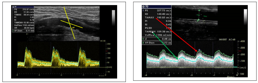
Figure 2. Flow volume measurement in the graft.
The flow volume calculation is based on the following formula: Qa 1⁄4 pr2 " TAMEAN, where r is the radius of the examined graft and TAMEAN is the time-averaged mean velocity inside. The latter is an integral value during the heart cycle of the average velocity layer (flow velocity is the fastest in the middle and the slowest along the vascular wall). TAMEAN must not be replaced by TAMAX, which is the time velocity integral of the fastest velocity only—using TAMAX would lead to significant overesti- mation of Qa. Pulse wave Doppler sample size should be wide enough to cover most of the vessel lumen because of the different speeds of blood inside the vessel from the center to the wall.
Figure 3. RD measurement.
There is excessive intimal hyperplasia in the outflow vein causing the stenosis. By diameter reduction, this stenosis would be significant if compared with the left or right part of the vein. Nevertheless, the RD is 2.35 mm, so this stenosis was considered borderline, and soon, reevaluation (within 6–8 weeks) was indicated.
ischemia.17 In considering preemptive correction, studies regarded stenoses as significant either at a flow volume decrease by 20%–25% and/or at a cut-off value of <500– 600 mL/min.13–14,18–20 However, the problem is that the actual Qa does not depend only on stenosis severity, but also on the actual hydration status and blood pressure similar to and in relation with the cardiac output. Natural/physiolo- gic variation of repeated Qa exceeds 20%.21 It is advisable to validate Qa values obtained by DUS with dilutional tech- niques performed during hemodialysis.
4
The Journal of Vascular Access
Residual diameter (RD) of the stenosis could be pre- cisely analyzed by the modern high quality ultrasound devices (Figure 3).8 The cut-off value <2.0 mm was arbi- trarily set and confirmed clinically when used as a part of the “complex stenosis criteria” in AVGs13,22,23 and vali- dated against angiography.8 In AVFs, an Australian study showed that the RD < 2.7 mm was associated with 90% sensitivity and 80% specificity of AVF dysfunction.18 According to a recent study, RD < 2.5 mm is associated with lower Qa.24 Another current Japanese study with AVFs determined the RD cut-off value to be <1.86 mm.14 As long as the stenosis is asymmetrical, it should be again visualized by two perpendicular views, and for the diagnosis of a significant stenosis, the RD should be below the cut-off value in both..
The resistive index (RI) of the flow pattern in the feed- ing artery has been tested in several studies, and although it is used in the assessment of the transplanted kidney,25 the results in AVFs and AVGs are not conclusive. Values above 0.6–0.7 indicated stenosis.26,27 The RI is defined by the following equation: RI 1⁄4 (PSV % end diastolic velocity)/PSV. RI increases especially in juxtaanastomotic stenoses.
Some centers and trials have used the complex criteria of a significant stenosis, comprising the combination of two main criteria (>50% diameter reduction þ PSR >3) and at least one additional criterion (RD, flow volume decrease, or low Qa—see Table 1).28 AVG stenoses with a lack of any additional criterion (1⁄4 borderline stenoses) possess low risk of thrombosis (ca. 1% during 6–8 weeks since the primary diagnosis).22 Only 54% of borderline AVF stenoses progressed into significant stenosis in the study by Castro et al.5 The complex stenosis criteria (Table 1) have been developed for the “Watch and wait strategy.” These criteria were used in the largest randomized trial testing ultrasound surveillance and proving its benefit,13 but also in other trials.19,28 Unfortunately, the majority of trials used only >50% diameter reduction as the only cri- terion of stenosis.9–11 The second largest randomized con- trolled trial (RCT) of ultrasound surveillance that did not prove its benefit used complex criteria, but included RD < 4.0 mm.29 The latter cut-off value is quite high and could be found in many normally functioning AVFs/AVGs.
The key question is this: can ultrasound surveillance provide reliable guidance for the clinician as to which ste- nosis should be treated and when? The identification of such high-risk stenosis is based on the understanding of stenosis pathophysiology and on their detailed morpholo- gical and functional (Qa) evaluation.22 DUS enables com- plex evaluation of the stenosis significance, which was, however, used only in a few published trials as mentioned above. Moreover, DUS performed early after AVG cre- ation can also predict future risk of AVG abandonment and thus select high-risk AVGs that could profit from the sur- veillance most.30 DUS is even more frequently performed
in AVFs after their creation, although the optimal criteria are discussed.31
AVF/AVG thrombosis, however, can also develop not only because of stenosis progression, but also due to other mechanisms, such as hypotension, dehydration, unwished outer compression during sleep, thrombophilia, and so forth. Moreover, the aforementioned complex stenosis cri- teria cannot be used mechanically. One such example is a branched AVF—if an outflow vein stenosis is diagnosed, but Qa (measured in the brachial artery) is adequate and the alternative outflow vein (branch) is suitable for hemodia- lysis needling, the stenosis could be left without interven- tion. Alternatively, when a decreased Qa is calculated, but there is no clear high-grade stenosis, it is worth to estimate patient’s hydration, for example, by the visualization of the inferior vena cava diameter and collapsibility when the ultrasound device is equipped with abdominal or echocar- diography probes.32 Whole-body bioimpedance spectro- scopy is frequently used in hemodialysis units for the assessment of hydration.
Stenoses in AVFs
Anatomically distal AVF is the first access of choice. The more frequent upper extremity fistulas include radio- cephalic, ulno-basilic and brachiocephalic fistulas, Gracz fistula (median cubital vein attached to the brachial or radial artery), and transposed basilic vein. They have a long life- span, low risk of complications, and their use is associated with the longest life expectancy.33 The main limitation of AVFs is non-maturation, as up to 40%–60% of AVFs are never suitable for hemodialysis without additional AVF pro- cedures to34 facilitate maturation and the maturation rate of AVFs is suggested as a surgical quality indicator.35 Many factors responsible for these unsatisfactory early results have been identified, and they lead to outflow vein or inflow artery stenoses. Early DUS can identify such stenosis and provide an indication to percutaneous or surgical therapy— so-called assisted maturation.36,37 Later on, stenoses develop mostly in the outflow vein close to the anastomosis in distal forearm AVFs and more proximally in the outflow vein (cephalic arch) in brachiocephalic AVFs.38
Stenoses in AVGs
The most frequent AVGs are forearm—they begin at the radial or brachial artery and are straight or curved and are attached to the basilic vein. More proximal grafts originate at the brachial or even subclavian artery and are attached to the cephalic or subclavian vein. Lower extremity grafts are created in the case of occluded central veins and are straight (between popliteal artery and some groin vein) or looped (femoral artery to femoral or great saphenous vein). AVGs have a higher rate of complications and a shorter lifespan than AVFs. Therefore, AVG is the vascular access
Malik et al.
5
that is created and recommended especially in patients with abandoned subcutaneous veins, with AVF non-maturation, but also in elderly patients, thanks to AVG’s higher maturation rate.39
Unfortunately, no currently available graft is as good as an AVF that matured without intervention. DUS performed early after AVG creation can predict future complications and thus select patients who would profit more from ultra- sound surveillance.30 AVGs are generally more prone to later stenosis development than AVFs. The typical site of stenosis development is at the venous anastomosis and at the adjacent part of the outflow vein. This is caused by the formation of intimal hyperplasia after creation. Cannulation-related stenoses could also develop in the graft itself, especially when the areal puncture technique is applied instead of the recommended rope-ladder tech- nique.40 In this case, the graft wall disruption and subse- quent healing are responsible. Both development of intimal hyperplasia and puncture healing are slow-acting mechan- isms, which give time for surveillance.22
Ultrasound surveillance in the guidelines
Some published vascular access guidelines support DUS as a screening tool for detecting stenosis and to perform pre- emptive interventions to prevent the loss of the AV access. The Kidney Disease Outcomes Quality Initiative (KDOQI) clinical practice guideline for vascular access, published in 2006, recommended direct flow measurement and DUS as preferred techniques that may be used in AVF surveillance and preemptive correction of luminal stenosis of >50% when the access flow rate is less than 600 mL/min in AVGs, and '400–500 mL/min in AVFs, even if the access is still able to provide adequate hemodialysis.41
Recent Spanish guidelines have recommended DUS as the first visualization method in the case of clinical suspi- cion on access dysfunction or stenosis, and regarding sur- veillance, both DUS and dilution methods for AVFs but not for AVGs.42 The European Society for Vascular Surgery guidelines also recommended DUS as a non-invasive tool to be the first line imaging method only in patients with suspected vascular access dysfunction.43 The new Eur- opean Renal Best Practice (ERBP) guideline on AV access specifies that the evidence for surveillance of AVFs is inconclusive and needs more research. In addition, they recommend not to perform routine surveillance of AVGs with access flow measurements or DUS.44
Clinical trials on vascular access surveillance
Current statements of the societies are based especially on the results of the metaanalyses.44,45 Their authors included all RCTs testing the benefit of DUS surveillance and pre- emptive treatment of stenoses. The conclusions of both
Table 2. The use of DUS as answered by the VAS council and board members.
Do you routinely perform/support ultrasound examination after access creation?
Do you perform/support duplex Doppler ultrasound in the case of access problems (puncture difficulties, limb edema, decrease of dialysis dose . . . ?)
Do you check the access by ultrasound regularly? Do you indicate PTA if a severe stenosis is found?
AVF AVG 75% 75%
100% 91%
55% 55% 73%a 73%a
PTA: percutaneous transluminal angioplasty; AVF: arteriovenous fistula; AVG: arteriovenous graft. Percentage of “YES” is depicted.
a Half responded “only if symptomatic.”
metaanalyses are similar: Preemptive stenosis correction of a functional arteriovenous access does not improve access longevity; although results for native AVF are pro- mising, existing evidence is insufficient to guide clinical practice and health policy.
The board and council members of the Vascular Access Society were asked about their practice in DUS surveil- lance. The results are given in Table 2, and they illustrate the current uncertainty about the clinical value of vascular access surveillance. The results of the largest RCT evi- denced the benefit of DUS surveillance and preemptive therapy of carefully assessed stenoses.13 On the contrary, the metaanalyses had negative results.45,46 Nevertheless, the number of patients included in the metaanalyses is scarce: six RCTs enrolled 612 patients with the primary endpoint of thrombosis, and four RCTs enrolled 443 patients for access loss.47 Some of the studies included prevalent patients, while others included solely incident patients at the time of access creation. The most important differences were, however, in the definition of significant stenosis that was indicated to percutaneous or surgical correction (Table 3). Performing a metaanalysis from so different studies leads therefore to significant oversimpli- fication. The final answer to whether to do or not to do DUS surveillance is therefore in including more patients with the use of the complex stenosis criteria and clear indication criteria of percutaneous treatment, ideally in a multicenter study with defined inclusion criteria and end- points (thrombosis and cumulative patency).
Perspective: is it the time to change the surveillance paradigm?
Although the guidelines are not favorable, the authors of this article are convinced that ultrasound should play a pivotal role in the AVF/AVG management, as it is a precise and highly reproducible method in experienced hands. For the greater distribution of AVF/AVG ultrasound imaging, it is necessary to (1) standardize the examination
6 The Journal of Vascular Access
Table 3. Duplex Doppler cut-off criteria of a significant AVG stenosis (used as the indication to angiography and intervention)—from the work by Malik et al.28
Trial first author
Mayer et al.9 Lumsden et al.10 Ram et al.11 Robbin et al.29 Malik et al.13
No of subjects US/control
35/35 32/32 32/34 65/61 97/92
Diameter reduction in B-mode
>50% >50% >50% >50% >50%
Peak velocity increase
Qa decrease
Qa cut-off value (mL/min)
Residual diameter (mm)
Prolonged1⁄4survival in ultrasound surveillance arm?
technique, (2) define the technical demands of the ultra- sound devices, and (3) standardize the criteria of signifi- cant stenosis in a well-controlled multicenter trial.
Ultrasound should be the first technique of choice in the evaluation of maturation, regardless of physical examina- tion. The goal in the maturation phase should be the search for potentially correcting inflow or outflow defects, often the basis of late stenoses. Hemodynamics and anatomy of AVF and graft vary from case to case, so the examination should be tailored for each patient in order to identify early the sites and causes of stenosis during the maturation phase. Although it has not been proven, it is logical that surveillance is most important in accesses at greater risk. To put things in perspective, top-end ultrasound devices and profound operator training should be a must in dedi- cated vascular access centers. Nevertheless, as in any field of medicine, clinical judgment is more important than only the results of ultrasonography. It includes also the history of previous thrombosis, thrombophilia, lack of other suit- able veins for another arteriovenous access, and so forth.
Declaration of conflicting interests
The author(s) declared no potential conflicts of interest with respect to the research, authorship, and/or publication of this article.
Funding
The author(s) disclosed receipt of the following financial support for the research, authorship, and/or publication of this article: JM is supported by the by the grant of the Agency of Health Research of the Czech Republic 17-31796A.
ORCID iDs
Jan Malik
Mario Meola
Cora de Bont
Joris I Rotmans
Jose Ibeas https://orcid.org/0000-0002-1292-7271
References
1. Tsai HM. Shear stress and von Willebrand factor in health and disease. Semin Thromb Hemost 2003; 29(5): 479–488.
https://orcid.org/0000-0002-2386-3293 https://orcid.org/0000-0002-1330-2820 https://orcid.org/0000-0003-2176-5275
– – – – Yes – – – – No – – – – No
>2fold >2fold
<500 <4.0 No >25% <600 <2.0 Yes
2. Remuzzi A and Ene-Iordache B. Novel paradigms for dia- lysis vascular access: upstream hemodynamics and vascular remodeling in dialysis access stenosis. Clin J Am Soc Nephrol 2013; 8(12): 2186–2193.
3. Maldonado-Carceles AB, Garcia-Medina J and Torres- Cantero AM. Performance of physical examination versus ultrasonography to detect stenosis in haemodialysis arterio- venous fistula. J Vasc Access 2017; 18: 30–34.
4. Malik J, Slavikova M, Malikova H, et al. Many clinically silent access stenoses can be identified by ultrasonography. J Nephrol 2002; 15(6): 661–665.
5. Castro A, Moreira C, Almeida P, et al. The role of Doppler ultrassonography in significant and borderline stenosis Def- inition. Blood Purif 2018; 46(2): 94–102.
6. Vachharajani TJ. Diagnosis of arteriovenous fistula dys- function. Semin Dial 2012; 25(4): 445–450.
7. Paulson WD. Blood flow surveillance of hemodialysis grafts and the dysfunction hypothesis. Semin Dial 2001; 14(3): 175–180.
8. Kudlicka J, Kavan J, Tuka V, et al. More precise diagnosis of access stenosis: ultrasonography versus angiography. J Vasc Access 2012; 13(3): 310–314.
9. Mayer DA, Zingale RG and Tsapogas MJ. Duplex scanning of expanded polytetrafluoroethylene dialysis shunts— impact on patient-management and graft-survival. Vascular Surg 1993; 27: 647–658.
10. Lumsden AB, MacDonald MJ, Kikeri D, et al. Prophylactic balloon angioplasty fails to prolong the patency of expanded polytetrafluoroethylene arteriovenous grafts: results of a prospective randomized study. J Vasc Surg 1997; 26(3): 382–390; discussion 390–392.
11. Ram SJ, Work J, Caldito GC, et al. A randomized controlled trial of blood flow and stenosis surveillance of hemodialysis grafts. Kidney Int 2003; 64(1): 272–280.
12. Robbin ML, Oser RF, Allon M, et al. Hemodialysis access graft stenosis: US detection. Radiology 1998; 208(3): 655–661.
13. Malik J, Slavikova M, Svobodova J, et al. Regular ultraso- nographic screening significantly prolongs patency of PTFE grafts. Kidney Int 2005; 67(4): 1554–1558.
14. Ishii T, Suzuki Y, Nakayama T, et al. Duplex ultrasound for the prediction of vascular events associated with
Qa 1⁄4 access flow volume.
Malik et al. 7
arteriovenous fistulas in hemodialysis patients. J Vasc
Access 2016; 17: 499–505.
29. Robbin ML, Oser RF, Lee JY, et al. Randomized compar- ison of ultrasound surveillance and clinical monitoring on arteriovenous graft outcomes. Kidney Int 2006; 69(4): 730–735.
30. Kudlicka J, Malik J, Tuka V, et al. Arteriovenous grafts: early ultrasonography tells their fortune. Am J Nephrol 2015; 41: 420–425
31. Lee T, Magill M, Burke SK, et al. Comparison of postopera- tive ultrasound criteria to predict unassisted use of arterio- venous fistulas for hemodialysis. J Vasc Access 2018; 19(2): 167–171.
32. Sabaghian T, Hajibaratali B and Samavat S. Which echo- cardiographic parameter is a better marker of volume status in hemodialysis patients? Ren Fail 2016; 38(10): 1659–1664.
33. Dhingra RK, Young EW, Hulbert-Shearon TE, et al. Type of vascular access and mortality in U.S. hemodialysis patients. Kidney Int 2001; 60(4): 1443–1451.
34. Dember LM, Beck GJ, Allon M, et al. Effect of clopidogrel on early failure of arteriovenous fistulas for hemodialysis: a randomized controlled trial. JAMA 2008; 299: 2164–2171.
35. Fila B, Roca- Tey R, Malik J, et al. Quality assessment of vascular access procedures for hemodialysis: a position paper of the Vascular Access Society based on the analysis of existing guidelines. J Vasc Access 2020; 21: 148–153.
36. Farrington CA, Robbin ML, Lee T, et al. Postoperative ultrasound, unassisted maturation, and subsequent primary patency of arteriovenous fistulas. Clin J Am Soc Nephrol 2018; 13: 1364–1372.
37. Abreo K, Buffington M and Sachdeva B. Angioplasty to promote arteriovenous fistula maturation and maintenance. J Vasc Access 2018; 19(4): 337–340.
38. Turmel-Rodrigues L, Pengloan J, Baudin S, et al. Treatment of stenosis and thrombosis in haemodialysis fistulas and grafts by interventional radiology. Nephrol Dial Transplant 2000; 15(12): 2029–2036.
39. Lee T, Qian J, Thamer M, et al. Tradeoffs in vascular access selection in elderly patients initiating hemodialysis with a catheter. Am J Kidney Dis 2018; 72(4): 509–518.
40. Parisotto MT, Schoder VU, Miriunis C, et al. Cannulation technique influences arteriovenous fistula and graft sur- vival. Kidney Int 2014; 86(4): 790–797.
41. Group VAW. Clinical practice guidelines for vascular access, update 2006—introduction. Am J Kidney Dis 2006; 48: S176–S247.
42. Roca-Tey R, Ibeas J, Moreno T, et al. Dialysis arteriove- nous access monitoring and surveillance according to the 2017 Spanish guidelines. J Vasc Access 2018; 19(5): 422–429.
43. Schmidli J, Widmer MK, Basile C, et al. Editor’s choice— vascular access: 2018 clinical practice guidelines of the European Society for Vascular Surgery (ESVS). Eur J Vasc Endovasc Surg 2018; 55(6): 757–818.
8
The Journal of Vascular Access
44.
45.
Gallieni M, Hollenbeck M, Inston N, et al. Clinical practice guideline on peri- and postoperative care of arteriovenous fistulas and grafts for haemodialysis in adults. Nephrol Dial Transpl 2019; 34: 1–42.
Tonelli M, James M, Wiebe N, et al. Ultrasound monitoring to detect access stenosis in hemodialysis patients: a sys- tematic review. Am J Kidney Dis 2008; 51: 630–640.
46. Ravani P, Quinn RR, Oliver MJ, et al. Preemptive correc- tion of arteriovenous access stenosis: a systematic review and meta-analysis of randomized controlled trials. Am J Kidney Dis 2016; 67: 446–460.
47. Tessitore N and Poli A. Pro: vascular access surveillance in mature fistulas: is it worthwhile? Nephrol Dial Transpl 2019; 34: 1102–1106.
Hai una richiesta o una domanda? Compila il nostro form per avere una risposta entro 24 ore.