Advances in vascular anatomy and pathophysiology using high resolution and multiparametric sonography
11297298211020150.pdf
Abstract
JVA The Journal of Vascular Access
B-mode and Color Doppler are the first-line imaging modalities in cardiovascular diseases. However, conventional ultrasound (US) provides a lower spatial and temporal resolution (70–100 frames per second) compared to ultrafast technology which acquires several thousand frames per second. Consequently, the multiparametric ultrafast platforms manage new imaging algorithms as high-frequency ultrasound, contrast-enhanced ultrasound, shear wave elastography, vector flow, and local pulse wave imaging. These advances allow better ultrasound performances, more detailed blood flow visualization and vessel walls’ characterization, and many future applications for vascular viscoelastic properties evaluation.
In this paper, we provide an overview of each new technique’s principles and concepts and the real or potential applications of these modalities on the study of the artery and venous anatomy and pathophysiology of the upper limb before and after creating a native or prosthetic arterio-venous fistula. In particular, we focus on high-frequency ultrasound that could predict cannulation readiness and its potential role in the venous valvular status evaluation before vascular access creation; on contrast-enhanced ultrasound that could improve the peri-operative imaging evaluation during US-guided angioplasty; on shear wave elastography and local pulse wave imaging that could evaluate preoperative vessels stiffness and their potential predictive role in vascular access failure; on vector flow imaging that could better characterize the different components of the vascular access complex flow.
Keywords
Multiparametric ultrasound, shear wave elastography, high-frequency ultrasound, vector flow imaging, local pulse wave velocity, vascular access
Date received: 19 June 2020; accepted: 6 May 2021
Introduction
Ultrasound (US) is the first-line imaging modality for screening, diagnosis, and monitoring treatment in cardio- circulatory pathology because of its safeness, non-inva- siveness, wide availability, and low cost. Conventional ultrasound modalities as B-mode, color Doppler, and spec- tral analysis allow the recognition of vessel wall and bloodstream changes, depicting the site of vascular sten- oses and occlusions.1,2 Consequently, US plays a pivotal role also in hemodialysis vascular access creation and surveillance.
Conventional platforms have a limited frame rate (images/second) because they use the line-by-line scanning acquisition method. This technical approach provides a lim- ited temporal resolution. Recent ultrafast platforms acquire
image information at frame rates of several thousand Hz, while conventional ultrasound systems acquire images at only 70–100 frames per second. The higher temporal and spatial resolution of this technical approach allows manag- ing all the new imaging algorithms as high-frequency
1Institute of Life Sciences, S. Anna School of Advanced Studies, Pisa, Italy
2Institute of Life Sciences, S. Anna School of Advanced Studies, Department of Internal Medicine, University of Pisa, Pisa, Italy
3Radiodiagnostic and Interventional Radiology Department, Fondazione IRCCS Policlinico San Matteo, Pavia, Italy
Corresponding author:
Petrucci Ilaria, Institute of Life Sciences, S. Anna School of Advanced Studies, Piazza Martiri della Libertà, 33, Pisa 56127, Italy.
Email: i.petrucci@santannapisa.it
The Journal of Vascular Access
1–8
© The Author(s) 2021
Article reuse guidelines: sagepub.com/journals-permissions hDttOpsI://1d0o.i1.o1r7g/71/01.1127977/1219289272191802210110250150 journals.sagepub.com/home/jva
2
The Journal of Vascular Access 00(0)
ultrasound (HFUS), contrast-enhanced ultrasound (CEUS), shear wave elastography, vector flow, and local pulse wave imaging. Ultrafast imaging allows better US performance, with more detailed visualization of blood flow, better char- acterization of vessel walls, and many applications in vascu- lar viscoelastic properties evaluation.
The availability of these technologies allows coining the term “high resolution” and “multiparametric ultra- sound” (MPUS) to define the current imaging applications on the top-line machine.3
Regarding vascular applications, the technological advances with potential applications in vascular access planning and surveillance are HFUS, CEUS, shear wave elastography, vector flow, and local pulse wave imaging.3–5
In this paper, we provide an overview of the principles and concepts of high resolution and MPUS, and we describe the actual or potential applications on the study of the artery and venous anatomy and pathophysiology of the upper limb before and after the creation of a native arterio- venous fistula (AVF) or a graft.
High-frequency ultrasound
High-frequency ultrasound refers to the US probe fre- quency of more than 10 MHz. High-frequency transducers were introduced in the pre-clinical setting and were mainly used in animal models to monitor tumor growth and to evaluate changes in micro-vascularization of neoplastic masses after antineoplastic therapy.4 The US transducers used in clinical practice operate at 22–100 MHz. These frequencies have short wavelengths and are more easily absorbed, so they improve spatial resolution with a reduced depth of penetration. HFUS could be applied to evaluate normal and diseased skin and superficial vessels.
Clinical applications
Nowadays, HFUS is used in neonatal and pediatric pathol- ogies to evaluate nerves, hand transplants, thyroid, lymph nodes, male reproductive organs, dermatological patholo- gies (melanoma, lipoma, hair follicles), and also musculo- skeletal and oral pathologies.6
The vascular applications of this technique are mainly on the smallest vascular anatomy: arteries and veins in pediatric and neonatal patients, measurement of Intima- Media Thickness (IMT) and characterization of carotid plaques for research and assessment of cardiovascular health, assessment of peripheral vessels in diabetes and other circulatory condition and visualization of flow pat- terns in atherosclerotic or abnormal vessels.
Vascular access applications
Jaberi et al.7 have hypothesized that venous wall thickness and circumferential stress measured with HFUS could
Figure 1. Cephalic vein valve and IMT evaluated with HFUS. Valve flaps are very well distinguishable (white arrows) and IMT (red line) is measurable at the far wall of the vessel.
predict cannulation readiness in AVFs. They have scanned an excised AVF specimen with a 55-MHz probe, and they have correlated the US findings with histological features. Then, they measured with HFUS the IMT of the near-field AVF venous wall of 20 patients with newly created AVFs within one week of cannulation. Cannulation readiness was defined as no extravasation during the first dialysis treatment. The mean IMT of the no extravasation group was greater than that of the extravasation group (p < 0.001). A minimum threshold IMT of 0.13 mm (p < 0.001) was associated with successful cannulation. The mean cir- cumferential stress of the no extravasation group was lower than that of the extravasation group (p < 0.001). A maximum circumferential stress threshold of 248 kPa was associated with successful cannulation (p = 0.009). They concluded that venous IMT and circumferential stress assessed with HFUS could predict cannulation readiness in AVFs clinically considered mature.
High-frequency US could also be useful in evaluating venous valves (Figure 1) during the preoperative mapping before AVF creation, and further studies are needed to evaluate its real utility.
Contrast-enhanced ultrasound
CEUS is an imaging modality based on microbubbles, made up of a hydrophilic shell surrounding a gas core, using their property of resonant volumetric oscillations in response to the acoustic pressure variations of the ultra- sound waves.
Ultrasound contrast enhancers are administered intra- venously in an aqueous solution as a bolus followed by a flush saline solution. Microbubbles have a mean size of 3 μm, and in general 95% of the bubbles pooling is smaller than 10 μm. They remain in the bloodstream, do not pass into the interstitial fluid and easily cross the pulmonary
Ilaria et al.
3
and peripheral capillaries without causing thrombotic complications. They are able to make “bright” the vessel, allowing an accurate study of the micro and macro-vascu- lature.8 Microbubbles are then disrupted by the acoustic US pulse and exhaled with breathing. CEUS imaging per- mits the evaluation of the vascular abnormalities, estimates blood flow parameters, and identifies and quantifies the neovascularization.9,10
Clinical applications
In literature, the use of CEUS has recently obtained many established and emerging applications.11 CEUS is rou- tinely employed for the differential diagnosis in focal parenchymal lesions, especially in the liver and in the kid- ney, for screening and early diagnosis of hepatocellular carcinoma in cirrhotic livers.12 Microbubbles allow to assess of the contrast enhancement due to malignant angi- ogenesis and identify the “wash-out” in the late phases.
CEUS imaging is also used to evaluate abdominal aortic aneurysm after endovascular aneurysm repair as an alterna- tive to computed tomography angiography, especially in contrast allergy or in patients with impaired renal function avoid excessive radiation exposure during follow-up.13 CEUS can accurately detect and characterize eventual endoleak, showing the persistence of contrast enhancement into the aneurysmal sac, which may lead to progressive aneurysmal enlargement and secondary rupture. CEUS is even superior to tomography angiography to detect the pres- ence and type of endoleaks in some cases. It is not affected by metallic artifacts of the graft, and it is a dynamic imaging modality allowing to visualize the inflow vessels source of endoleak. Limitations are similar to conventional US imag- ing: operator dependence and image quality impairment depending on body habitus and bowel gas presence.14 The risk of anaphylactic reaction after ultrasound contrast agent administration is very rare (1 on 100.000 cases).
CEUS imaging is also spreading in the Emergency Department, where this method permits the identification of aortic dissection and post-traumatic parenchymal inju- ries and finds eventual focal sources of bleeding.15
CEUS imaging allows an accurate measurement of the degree and length of stenosis and better evaluates plaque morphology, thickness, and vulnerability. For these rea- sons, many studies applied this imaging modality to assess the extracranial carotid occlusive disease and estimate the carotid plaque. It was demonstrated the possibility to detect and quantify intraplaque angiogenesis, which is a marker of plaque growth.16 Thus, CEUS may be used for risk stratification of patients with atherosclerotic disease and predict the risk of cerebrovascular accident.
Vascular access applications
Ramnarine et al.17 reported the use of CEUS in 11 patients in ultrasound guided AVFs balloon angioplasty procedures
for failing or non-maturing AVFs. The procedure was under the guidance, and CEUS was used initially to evalu- ate the fistula morphology and after balloon angioplasty to confirm the stenosis treatment and reveal intra-operative complications. They conclude that CEUS improves peri- operative imaging evaluation because it provides a higher spatial resolution for narrow stenotic segments. It is more sensitive in demonstrating potential complicating extrava- sation, avoiding the use of iodinated contrast, and being a viablealternativetofluoroscopyforAVFintervention.The main limitations are that more proximal lesions may be difficult to visualize due to depth limitation.
Tissue elastography
Mechanical tissue properties change in many different dis- ease processes that lead to fibrosis, inflammation, and neo- vascularization. US elastography evaluates the tissue stiffness based on Young’s modulus, a physical property that relates applied force per unit area (stress) and the conse- quent relative change in tissue dimension (strain). Ultrasonographic methods to evaluate tissue elastography may be strain-based, in which the probe pressure applies the force, or shear wave-based, in which the force is produced by the imaging system.18 Strain elastography only allows semi-quantitative assessments of stiffness that are difficult to compare longitudinally. Shear wave propagation velocity is directly correlated with tissue stiffness, and in most ultra- sound systems, compressive acoustic waves are used to induce and track shear waves all along with the ultrasound probe, permitting shear wave velocity estimation.
US elastography has been successfully applied to eval- uate many tissues and organs, including liver, breast, thy- roid, kidney, spleen, prostate, lymph nodes, tendons, and vessels.18,19
Clinical applications
Strain elastography is an accurate technique to distinguish liver fibrosis stages and benign from malignant liver masses. Shear wave elastography (SWE) is used since the early stages of liver fibrosis caused by HBV, HCV, alco- holic liver disease, hepatic toxicity, and autoimmune hepa- titis. In patients with chronic kidney disease (CKD), strain values are higher than in healthy volunteers, and strain elastography can also detect early renal graft interstitial fibrosis, suggesting an organ rejection. SWE has shown significant differences between CKD grades, and it could also represent a marker for diabetic kidney disease.18 Moreover, strain-imaging studies to assess focal renal masses have shown promising results. SWE has improved the differential diagnosis between benign and malignant breast, thyroid, pancreas, and prostate lesions, and encour- aging results are growing about the use of SWE in the dif- ferential diagnosis between benign and malignant lymph nodes, endoscopic spleen evaluation in chronic liver
4
The Journal of Vascular Access 00(0)
disease, and cirrhosis.19 The cardiovascular system is a new potential field of application of SWE, especially in the risk stratification of carotid plaques3 and in the evalua- tion of carotid stiffness in patients with acute ischemic stroke.20 Arterial stiffness measured with SWE has been evaluated in patients with CKD, and the results show that their brachial artery is stiffer than in healthy subjects.21 At present, there is only limited evidence available for the application of SWE in the evaluation of peripheral veins, in particular on thrombus characterization.3
Vascular access applications
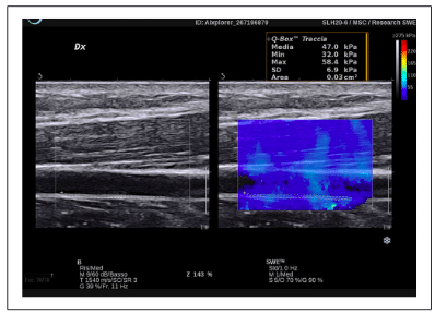
Inabilities to prevent non-maturation or avoid early fail- ure of AVFs are due to an incomplete understanding of preexisting arterial and venous conditions. Small-diameter at anastomosis sites, old age, diabetes mellitus, arterio- sclerotic pathologies of the artery, and preexisting low- quality vein wall are all risk factors of non-maturation and early failure of vascular access.22 Whether preexisting vascular pathologies associated with or aggravated by CKD can impair the necessary adaptive remodeling of blood vessels after vascular access placement is still an open question. There is some evidence that the elastic modulus measurements are significantly higher in patients undergoing preoperative mapping for hemodialysis access than in healthy volunteers.23 The arterial stiffness could be a possible biomarker for AVF failure due to the lower arterial ability to dilate.24–26 However, the studies pub- lished to date on the arterial stiffness evaluation before or after AVFs creation are not conclusive because they have used very heterogeneous methods, not including SWE that could potentially be able to evaluate the local arterial stiffness.27–29 MacDonald et al.30 have retrospectively analyzed the preoperative and postoperative data of 33 patients indicated for fistula creation. Vessels diameters at the B-mode ultrasound, SWE of the brachial artery, and demographic data were considered to find if any of these variables were related to the outcome of the AVF 3 months after creation. Shear wave velocity decreased after AVF creation, indicating increased compliance, but there were no parameters associated with AVF failure. Further stud- ies with a larger number of patients and a longer follow- up period are needed to assess the utility of SWE evaluation of arterial and or venous vessels before and after AVF (Figure 2) or AVG creation (Figure 3).
Vector flow imaging
Vector Flow Imaging (VFI) is an innovative imaging algo- rithm, angle-independent, which provides a multidimen- sional characterization of blood flow in all directions, showing the streamlines and vortices distribution into a vessel as velocity vectors.31
Figure 2. Longitudinal view of a cephalic vein in a distal radio-cephalic AVF using local SWE. The region of interest is adjusted to the IMT of the far wall of the vein. In this case the venous IMT is homogeneous with a stiffness of 47 kPa.
Figure 3. Longitudinal view of the venous anastomosis in a prosthetic arteriovenous fistula using SWE, which shows the different elastic properties and stiffness of the vessel walls. The portion of the graft is much stiffer (represented in red) at the venous anastomosis than the contiguous venous walls.
Commercial US systems primarily developed two methods of estimation based on different principles: trans- verse oscillation method and plane wave imaging. In the transverse oscillation method, the probe emits several con- secutive pulses. Two receiving beamformers are used to estimate the axial and transverse components of the veloc- ity vectors, and the difference from the received signals is compared to calculate the velocity.32,33
In ultrafast plane wave imaging, the transmission is made up of single unfocused beams at several incidence angles. The multidirectional flow estimation is performed through speckle tracking, and the real vector velocity is calculated by compounding velocities returning from a series of steers through a complex angle-compounding algorithm.34–36
Ilaria et al.
5
These methods allow a high frame rate, with a dynami- cal and intuitive visualization of complex hemodynamic features. The bloodstream is represented in VFI with many moving-colored arrows, where the color and the length of the vector indicate the velocity magnitude.37 The operator can measure the velocity at any point of the vessel and every instant of the cardiac cycle, revealing even transient flow movements, otherwise not detectable with conven- tional ultrasound.
VFI has some limitations. It is based on pulse repetition frequencies, so it does not permit the correct measure- ments of higher velocities in aliasing areas, and it is a two- dimensional technique, not allowing a 3-D comprehension of the streamlines.
Clinical applications
Several clinical studies based on VFI showed the additional information carried out from this technique analyzing differ- ent flow patterns in the human cardio-circulatory system.
The first in-vivo study with the vector velocity method, based on plane wave acquisition, was published in 2009 by Hansen et al.,38 which reported the visualization of a stable vortex in the bulb of six carotid bifurcations.
VFI was then deployed to demonstrate reversal blood flow from the external to the internal carotid during the sys- tole, suggesting the possible role of retrograde embolism in the case of plaques in the proximal external carotid artery.39
Goddi et al.37 evaluated 60 carotid bifurcations in healthy adults, identified a complex flow in the internal carotid artery, and analyzed the location and the duration of the different blood flow patterns, confirming the rela- tionship between vessel enlargement and flow distur- bances. VFI was also used to assess the normal values of wall shear stress in the common carotid arteries of 79 healthy volunteers, with a good intraclass correlation coef- ficient and inter-observer reproducibility.40
Hansen et al.41 demonstrated the association between the vector concentration in VFI and the stenosis degree percent- age obtained with digital subtraction angiography in 11 patients with stenoses of the superficial femoral artery.
In a rare case of the femoral artery “trifurcation,” where the origin of the lateral circumflex femoral artery arises from the femoral bifurcation, VFI revealed a blood recir- culation rest and retrograde blood flow redistribution dur- ing lower limb compression.42
These preliminary evaluations demonstrate that high frame rate VFI represents an innovative ultrasound appli- cation, with a better and more intuitive flow estimation within the vessel during the cardiac cycle.
Vascular access applications
Hansen et al.43 compared the blood flow volume meas- ured during dialysis sessions with VFI and the ultrasound
Figure 4. Longitudinal view of a radio-cephalic fistula
using VFI, which represents the flow with many colored vectors frame. In this frame at the systolic peak it shows high velocity red vectors at the arterial side just before the fistula anastomosis, with recirculation and reverse flow (1), multidirectional low-velocity green vector against the venous wall on the venous side of the anastomosis (3) and faster vector streamline at the venous side of the fistula (2).
dilution technique in 20 AVFs, evidencing a good corre- lation with these methods. The possible sources of error during the volume flow rate measurement with VFI are related to the off-axis placement of the scan plane com- pared to the vessel’s central axis.44
A recent study reported the use of VFI in 14 AVFs to identify the different components that compound a com- plex flow (Figure 4). The authors found the presence of a disturbed flow, probably related to oscillatory wall shear stress and neointimal hyperplasia development, at the inner wall of the juxta-anastomotic venous side, into the venous aneurysmal tracts and in concomitance of stenosis.45
Pulse wave velocity measurement
Arterial stiffness is a predisposing factor for peripheral vascular disease and represents, in atherosclerosis, an independent risk marker for cardiovascular disease.46 Arterial stiffness can be measured with many different methods, but pulse wave velocity (PWV) is considered the most reliable. PWV is the velocity at which the blood pres- sure pulse propagates through the circulatory system.47 The carotid-femoral PWV is accepted as the standard for measuring aortic stiffness based on a “propagation model” of the arterial tree. However, it has several limitations because it needs a dedicated device, the measurement is not easily feasible and repeatable, and, finally, it does not assess a local arterial stiffness difference.48 Two novel techniques are currently being evaluated for PWV meas- urement: SWE, treated in a previous section, and ultrafast ultrasound imaging or UltraFastEcho (or UltraFast® Imaging), which measures the local PWV at the beginning
6
The Journal of Vascular Access 00(0)
and the end of systole.48 The technological innovation is based on the extremely high imaging frame rate, which is one hundred times faster than conventional ultrasound diagnostic imaging, thus capturing the propagation of the pulse wave in a localized segment of an artery during a single cardiac cycle. The evaluation of local PWV is becoming an important tool because local PWV has emerged as a powerful independent predictor of all-cause and cardiovascular mortalities.5
Clinical applications
Many studies in the literature have proven that global PWV is a predictor of all causes and cardiovascular causes of death in the general population.49 In CKD, the arterial stiffness measured with global PWV is worse in diabetic compared with no diabetic patients. PWV worsens as kid- ney function declines irrespective of the cause of CKD. It correlates with proteinuria in CKD diabetic patients and bone and mineral disorders. PWV is associated with higher central pulse pressures and predicts the onset of heart fail- ure, death, and CKD progression to end-stage renal dis- ease.50 Currently, local PWV measurements are being studied in order to provide localized information on arte- rial stiffness in many fields, such as vessel elasticity abnor- malities of individual target arteries and the prediction of cardiovascular events and end-organ damage, classifica- tion of normal and pathological arteries in hypertensive patients, assessment of coronary artery hemodynamics and determination of acute coronary events, non-invasive assessment of fetal hemodynamics, characterization of the retinal and ocular circulation analysis, and assessment of microvascular stiffness.
Vascular access applications
The role of PWV measurement in vascular access evalua- tion is not encouraging in literature. In the Hemodialysis Fistula Maturation Study on 602 patients undergoing AVF creation, Dember et al.29 that there is a low correlation between global PWV measurement and demographic, clinical, and biochemical factors among the different vas- cular function measures. Allon et al.,51 in the same group of patients, have stated that despite the hypothesis for which the stiffness of the arterial conduit used to create the AVF would restrict arterial outward remodeling, the study failed to find such a relationship. The carotid-femoral PWV showed an inverse relationship with 6-week AVF diameter, but there were no statistically significant rela- tionships between carotid-femoral PWV/carotid-radial PWV and AVF blood flow. Masengu et al.26 and McGrogan et al.52 did not find any statistically significant association between carotid-femoral PWV, brachial-radial PWV, and AVF early failure. However, in the second study, the aortic
Figure 5. Ultrafast measurement of the local PWV of the brachial artery. Ultrafast PWV measurements are obtained at the beginning of systole (BS) and the end of systole (ES). The region of interest is adjusted to the entire field of view of the transducer.
PWV was slower in the primary patency group than the primary failure group, suggesting that stiffer vessels lead to a higher probability of AVF primary failure.
In any case, in all studies, the techniques used for PWV evaluation represent indirect measures of arterial stiffness and are influenced by the global evaluation of several different arteries that may differ in their stiffness. Consequently, local PWV measurement could represent a direct evaluation of brachial and radial artery stiffness (Figure 5). For this reason, it would be very interesting to perform studies on the preoperative evaluation of local arterial stiffness before AVF creation and the correlation with AVF outcomes.
Conclusion
The technological advances encompassed in MPUS have many potential and interesting applications in vascular access planning and surveillance, and only a few of them have already been successfully applied. However, studies with many patients are needed to standardize the parame- ters for the study of the upper limb vascular anatomy and physiopathology before and after AVF/AVG creation and to evaluate their potential correlation with vascular access outcomes.
Declaration of conflicting interests
The author(s) declared no potential conflicts of interest with respect to the research, authorship, and/or publication of this article.
Funding
The author(s) received no financial support for the research, authorship, and/or publication of this article.
Ilaria et al.
7
ORCID iD
Petrucci Ilaria
References
17. Ramnarine R, Freedman B, Navaratne S, et al. Novel use of contrast enhanced ultrasound (CEUS) in angioplasty of hae- modialysis arteriovenous fistulas: initial experience. ECR 2013; C–2530.
18. Ozturk A, Grajo JR, Dhyani M, et al. Principles of ultra- sound elastography. Abdom Radiol 2018; 43: 773–785.
19. Sigrist RMS, Liau J, El Kaffas A, et al. Ultrasound elas-
tography: review of techniques and clinical applications.
Theranostics 2017; 7: 1303–1329.
20. Li Z, Du L, Wang F, et al. Assessment of the arterial stiff-
ness in patients with acute ischemic stroke using longitu- dinal elasticity modulus measurements obtained with shear wave elastography. Med Ultrason 2016; 18: 182–189.
21. MacDonald C, Ross R and Houston JG. Shear wave veloc- ity measurements of the brachial artery in a population with end-stage renal disease. Biomed Phys Eng Express 2018; 4: 057002.
22. Vazquez-Padron RI and Allon M. New insights into dialysis vascular access: impact of preexisting arterial and venous pathology on AVF and AVG outcomes. Clin J Am Soc Nephrol 2016; 11: 1495–1503.
23. Sorace AG, Robbin ML, Umphrey H, et al. Ultrasound measurement of brachial artery elasticity prior to hemodi- alysis access placement: a pilot study. J Ultrasound Med 2012; 31(10): 1581–1588.
24. McGrogan DG, Maxwell AP, Khawaja AZ, et al. Current tools for prediction of arteriovenous fistula outcomes. Clin Kidney J 2015; 8: 282–289.
25. Kheda MF, Brenner LE, Patel MJ, et al. Influence of arte- rial elasticity and vessel dilatation on arteriovenous fistula maturation: a prospective cohort study. Nephrol Dial Trans 2010; 25: 525–531.
26. Masengu A, Hanko JB and Maxwel AP. Arterial stiffness and arteriovenous fistula failure of maturation. J Vasc Access 2016; 17: 477–482.
27. Weitzel WF, Kim K, Park DW, et al. High-resolution ultra- sound elasticity imaging to evaluate dialysis fistula stenosis. Semin Dial 2009; 22: 84–89.
28. Paulson WD. Does vascular elasticity affect arteriovenous fistula maturation? Open Urol Nephrol J 2014; 7: 26–32.
29. Dember LM, Imrey PB, Duess MA, et al. Vascular function at baseline in the hemodialysis fistula maturation study. J Am Heart Assoc 2016; 5(7): e003227.
30. MacDonald CJ, Ross R and Houston JG. Increased brachial artery compliance after arteriovenous fistula creation as measured by shear wave elastography. J Ultrasound Med 2020; 39(5): 875–881.
31. Goddi A, Bortolotto C, Fiorina I, et al. High-frame rate vec- tor flow imaging of the carotid bifurcation. Insights Imaging 2017; 8: 319–328.
32. Udesen J and Jensen JA. Investigations of transverse oscilla- tion methos. IEEE Trans Ultrason Ferroelectr Freq Control 2006; 53: 959–971.
33. Udesen J, Nielsen MB, Nielsen KR, et al. Examples of in vivo blood vector velocity estimation. Ultrasound Med Biol 2007; 33: 541–548.
34. Yiu BY, Lai SS and Yu AC. Vector projectile imaging: time-resolved dynamic visualization of complex flow pat- terns. Ultrasound Med Biol 2014; 40: 2295–2309.
35. Ekroll IK, Dahl T, Torp H, et al. Combined vector veloc- ity and spectral Doppler imaging for improved imaging of
8
The Journal of Vascular Access 00(0)
complex blood flow in the carotid arteries. Ultrasound Med
Biol 2014; 40: 1629–1640.
44. Jensen J, Olesen JB, Stuart MB, et al. Vector velocity volume flow estimation: sources of error and corrections applied for arteriovenous fistulas. Ultrasonics 2016; 70: 136–146.
45. Fiorina I, Raciti MV, Goddi A, et al. Ultrasound vector flow imaging – could be a new tool in evaluation of arteriovenous fistulas for hemodialysis? J Vasc Access 2017; 8: 284–289.
46. Van Bortel LM, Laurent S, Boutouyrie P, et al. Expert con- sensus document on the measurement of aortic stiffness in daily practice using carotid- femoral pulse wave velocity. J Hypertens 2012; 30: 445–448.
47. Luo J, Li RX and Konofagou EE. Pulse wave imaging of the human carotid artery: an in vivo feasibility study. IEEE Trans Ultrason Ferroelectr Freq Control 2012; 59: 174–181.
48. Messas E, Pernotb M and Couade M. Arterial wall elas- ticity: state of the art and future prospects. Diagn Interv Imaging 2013; 94: 561–569.
49. Ben-Shlomo Y, Spears M, Boustred C, et al. Aortic pulse wave velocity improves cardiovascular event prediction: an individual participant meta-analysis of prospective observa- tional data from 17,635 subjects. J Am Coll Cardiol 2014; 63: 636–646.
50. Townsend RR. Arterial stiffness in CKD: a review. Am J Kidney Dis 2018; 73: 240–247.
51. Allon M, Greene T, Dember LM, et al. Hemodialysis Fistula Maturation Study Group. Association between preoperative vascular function and postoperative arteriovenous fistula development. J Am Soc Nephrol 2016; 27: 3788–3795.
52. McGrogan DG, Stringer S, Cockwell P, et al. Arterial stiff- ness alone does not explain arteriovenous fistula outcomes. J Vasc Access 2018; 19: 63–68.
Hai una richiesta o una domanda? Compila il nostro form per avere una risposta entro 24 ore.